Applications of Polypeptides in Biology
Diblock Copolypeptide Hydrogels (DCH) for Central Nervous System Repair

brain2.png
scaffolds2.png
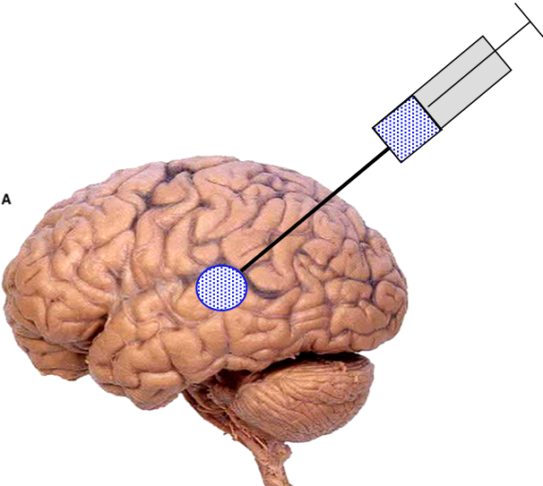
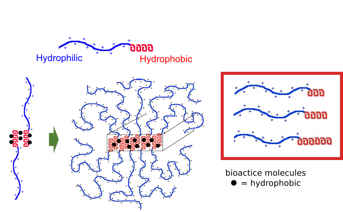
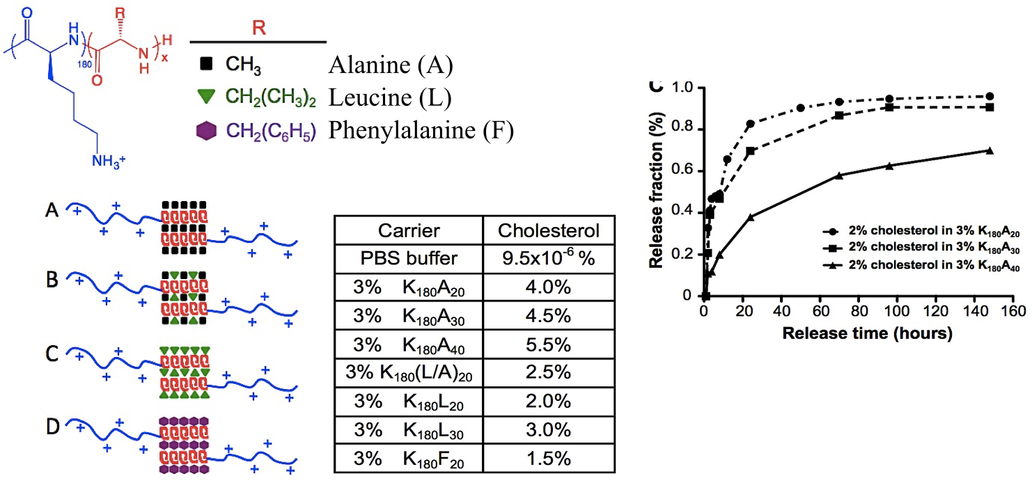
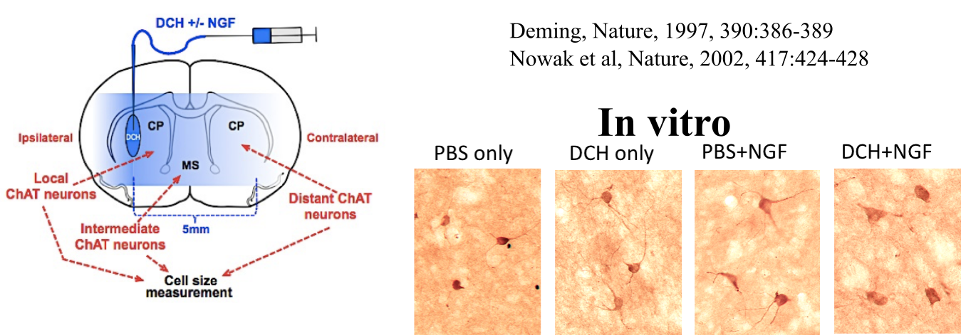
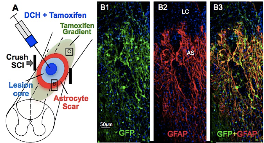
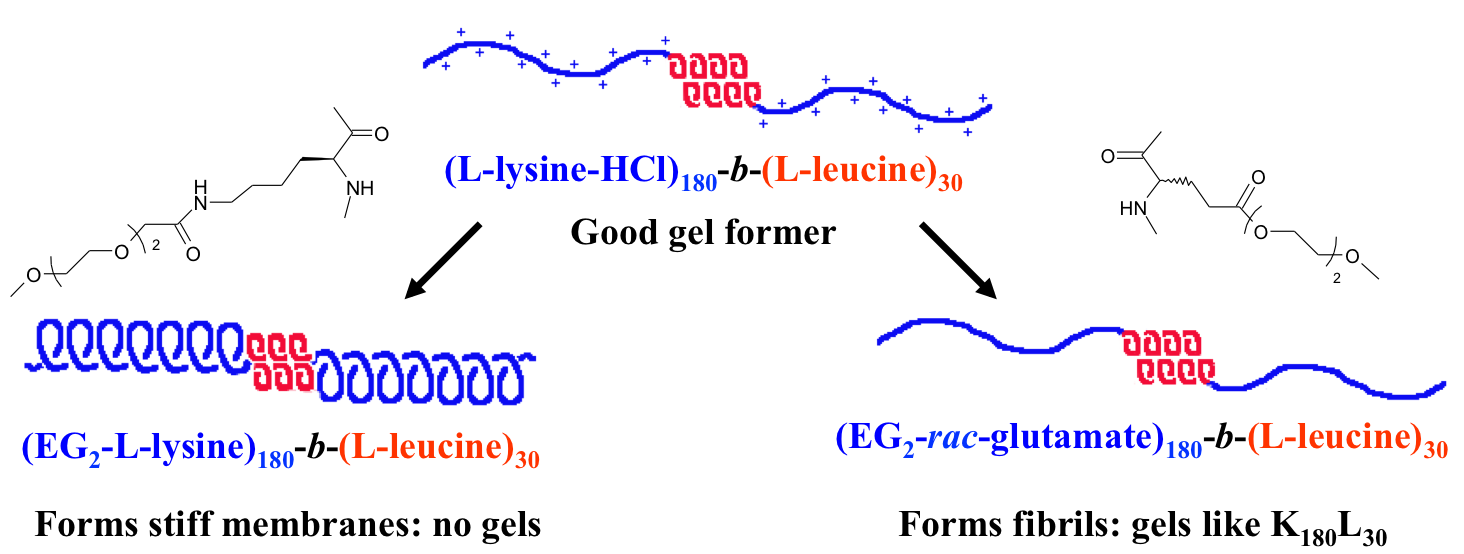
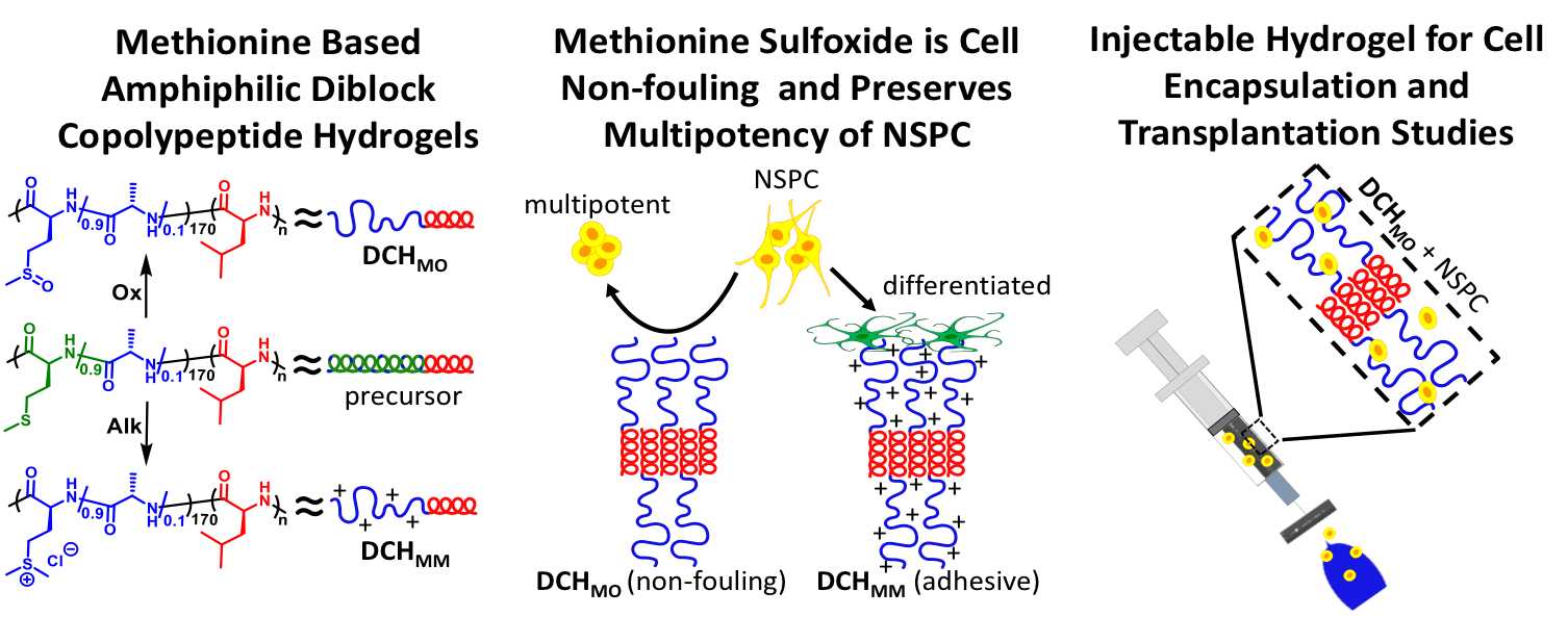
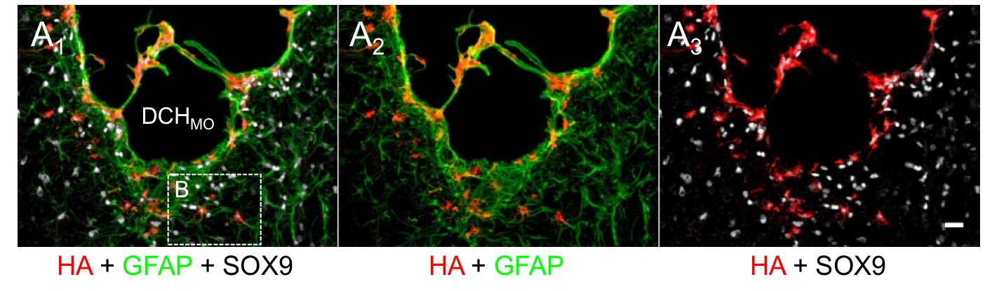
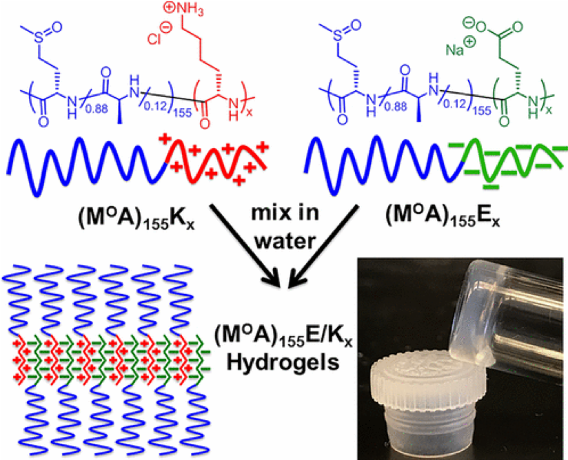
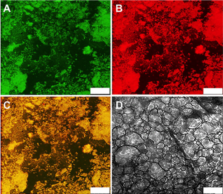
In collaboration with the lab of Prof. Michael Sofroniew (Neurobiology Dept, UCLA), were are developing polypeptide hydrogels for study of biology and neural repair in central nervous system (CNS) tissues. Synthetic DCH have been designed with tunable physical properties (stiffness, porosity, chemical functionality), and can be degraded in vivo. We have shown that many DCH formulations form hydrogel deposits and are well tolerated in healthy mouse forebrain tissue. DCH are being developed to encapsulate and release both hydrophilic (e.g. protein) and hydrophobic (e.g. small molecule active agent) cargos within CNS tissues. We are also developing DCH formulations to encapsulate neural progenitor stem cells (NPSCs) to allow cell grafting within CNS tissues, and to provide biomimetic scaffolds for encapsulated cells. Recent efforts have focused on study and development of DCH formulations to facilitate neural repair after spinal cord injury.
Encapsulation of Hydrophobic Molecules within DCH

hydrophobicLoading.png
The hydrophobic domains of DCH are able to dissolve small hydrophobic molecules. These can be small molecule drugs or signalling molecules.
Tuning of Cargo Loading Capacity and Release Rate

Loadingcapacity.png
Variation of hydrophobic segments in DCH allows adjustment of loading capacity and release rate
Encapsulation and Release of Growth Factors (proteins) in DCH

DCHgrowthfactor.png
Release of nerve growth factor (NGF) from a DCH depot in healthy mouse forebrain shows biological effects on cholinergic neurons (ChAT) in vivo, where they respond to NGF by increasing in size. DCH provide prolonged release of NGF compared to injection of NGF in saline.
Encapsulation and Release of Small Molecules in DCH

DCHgrowthfactor1.png
Release of Tamoxifen from a DCH depot in the lesion core of a spinal cord injury model in mouse shows biological effects on scar forming astrocytes. In the astrocytes (also identified using GFAP), Tamoxifen activates reporter gene expression of green fluorescent protein (GFP) in transgenic mice.
Non-Ionic DCH for Cell Grafting

nonionichydrogels.png
To improve cell compatibility, non-ionic DCH were developed. In the initial design, oligoethylene glycol functionalized polypeptide segments were used to replace cationic lysine segments found in original DCH. These DCH were able to support encapsulated NPSCs, which could then be grafted into CNS tissue with high viability and integration with host tissue.
Poly(Methionine Sulfoxide) based Non-Ionic DCH

MOXhydrogel.png MOXhydrogel2.png
As we continue to develop and optimize our DCH formulations, we have switched from unnatural oligoethylene glycol based hydrophilic segments in non-ionic DCH to use of natural poly(methionine sulfoxide) segments (DCHMO). The sulfoxide functionality in these hydrogels possesses non-fouling properties (similar to PEG), but gives DCH that are degradable and resorbable, in addition to being less expensive and easier to synthesize. DCHMO is currently being used for a variety of NPSC grafting studies in mice.
Polyion Complex DCH (DCHPIC)

PIChydrogel.png PIChydrogel1.png
Another recent development has been the design of DCH that can assemble via polyion complexation as opposed to hydrophobic interactions. By taking advantage of the ability of enantiomerically pure poly(lysine) and poly(glutamate) segments to form beta-sheet structured polyion complexes, we were able to form DCH that retain the functionality of DCHMO, but are assembled via stronger electrostatic and H-bonding interactions. The result is DCHPIC that are significantly more resistant to dissolution compared to DCHMO, and can be prepared with much greater stiffness as two-component formulations while retaining cell and tissue compatibility.